Scanning the Surface of an Unknown World: Studying Leaf Trichomes in the Ironweeds (Vernonieae, Compositae)
By Jackie Martinez
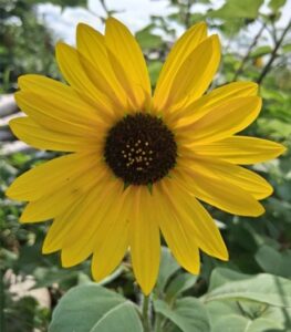
Figure 1. Sunflowers are members of the Compositae family aka Asteraceae. Photo courtesy Jackie Martinez.
Did you know that asters, daisies, sunflowers, and ironweeds are all part of the same plant family known as Compositae (Fig. 1)? In fact, Compositae (also referred to as Asteraceae) is believed to be THE largest family of flowering plants with over 33,000 species- accounting for nearly one in every 10 species of flowering plants! This astounding abundance of species has made the classification of Compositae a complex feat. A team of talented botanists from the Fort Worth Botanic Garden | Botanical Research Institute of Texas (FWBG | BRIT), Morris Arboretum and Gardens of the University of Pennsylvania, and Royal Botanical Gardens, Kew have come together to work towards reclassifying over 700 species, originating from the Eastern Hemisphere, of the “ironweed” tribe (also known as Vernonieae) in the flowering plant family, Compositae. This work has been made possible through a grant from the National Science Foundation (NSF) and Natural Environment Research Council (NERC), which has also sponsored my participation in this project through this internship.
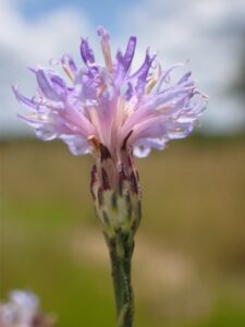
Figure 2. Linzia glabra is a species of ironweed formerly known as Vernonia glabra. Image courtesy M. Gostel.
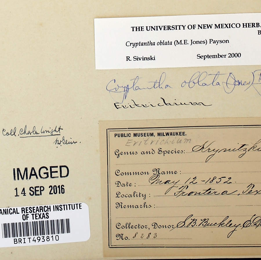
When I applied for the Botanical Science Internship at BRIT, I was looking forward to an experience that would enlighten me on the work that goes into botanical research, and I feel privileged to have had this exceptional opportunity. I learned so much from my mentors, Post Doctoral Researcher, Dr. Oyetola Oyebanji, and PhD. Candidate, Mihaja Andriamanohera based at the University of Texas at Arlington and working under the guidance of Principal Investigator, Dr. Morgan Gostel on better understanding plant diversity in the ironweeds. I began my work at the BRIT Philecology Herbarium collecting samples of various plant structures from several herbarium specimens from the genera Gymnanthemum and Vernonia (Fig. 2). Mihaja explained how to interpret the unique information found on the label of each specimen identifying the collector(s), the individual credited for naming the species, the country and coordinates from which it was collected, as well as additional identifying information (Figs. 3 & 4). Mihaja also stressed the importance of delicately collecting only as much material as needed for analyses, as it is important to preserve all herbarium specimens as irreplaceable and permanent treasures that will continue to inform biodiversity research in years ahead.
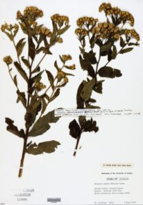
Figure 3. Herbarium voucher of Linzia glabra. Image © University of Georgia Herbarium.
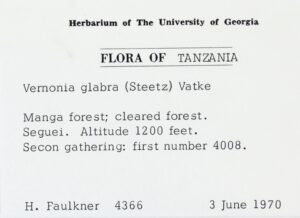
Figure 4. Label for Linzia glabra including identifying information on the specimen. Image © University of Georgia Herbarium.
After collecting a set of samples, I spent time in the dry lab where Oyetola and Mihaja coached me on properly preparing the collected material for scanning electron microscope (SEM) imaging. The SEM is a microscope capable of obtaining high-resolution images of objects invisible to the naked eye at remarkably high-magnification, which allows the observer to observe and record key information to compare and describe species and their structures. These structures include tiny details like pollen, hairs or outgrowths on the surface of plants (called trichomes), and the distribution of microscopic pores on the surface of leaves that allow plants to “breathe” (called “stomata”). I had the pleasure of being trained on operating the SEM by Marsha Stripe, an established and highly knowledgeable volunteer at BRIT. Marsha made sure to set me up for success; she explained how the instrument worked, what protocol to follow when mounting and inserting the samples into the chamber, and how to fine-tune a scan to capture high-quality images. I felt very confident in myself by the time my training was complete (Fig. 5).
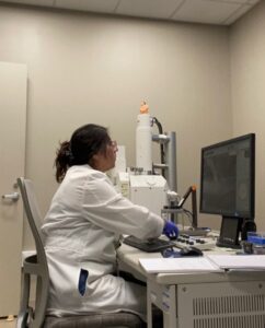
Figure 5. Spring intern Jackie Martinez viewing Compositae leaf samples in the BRIT SEM lab. Photo courtesy Martha Stripe.
The SEM lab is where the magic happened for me and the material I had so meticulously collected and prepared came to life! I was fascinated by the distinct micromorphological variation I saw across species- even within the same species (Fig. 6 and 7). Some of the leaf surfaces I observed were barren like the surface of Mars with only the presence of stubs from broken off trichomes and shriveled glandular trichomes. Others were so incredibly dense with trichomes you couldn’t even see through to the surface of the leaf – like the plant leaves were wearing a thick winter coat. Some species had stomata present on both the adaxial (upperside) and abaxial (underside) axes of the leaf while most of the samples I viewed had them exclusively on the abaxial axis.
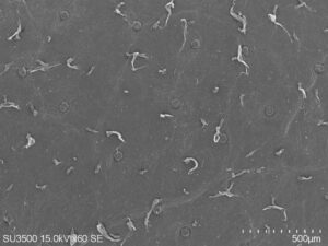
Figure 6. SEM image of sample T13. View of the adaxial leaf surface of Gymnanthemum exsertiflorum. Image courtesy Jackie Martinez.
Observing, recording, and documenting these physical characters is only a piece of the puzzle to understanding the evolutionary relationships of the hundreds of species of ironweeds worldwide, but the micromorphological features found on these individual species will contribute significant data to support a revised subtribal classification (this is technical “taxonomic speak” for determining how to accurately classify and name species in this group). We will use these data, together with DNA sequence data to build something similar to a family tree (called a “phylogenetic tree”) to understand how these species are related to one another, how they have changed over millions of years, and describe potential new species, I am fortunate to have played a role in this multifaceted and ongoing study, and I am grateful to all the people I met and worked with during my time at BRIT.
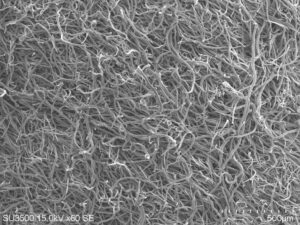
Figure 7. SEM image of sample T14. View of the abaxial leaf surface of Gymnanthemum exsertiflorum. Image courtesy Jackie Martinez.
BRIT Experience Blog
Summer Internship
This article was written by Spring 2026 NSF-funded Ironweeds Intern, Jackie Martinez.